Posted by Gregory J Block MSc PhD on Jan 11, 2015

You may also know that this is a relatively fresh consensus, but that over the last two years, our funding has already screened over 3 million compounds looking for inhibitors of DUX4. Screening for inhibitors is great. But what about when you find something that works, and you need to tweak it a little bit here and a little bit there?? Or what if you want to design something from the ground up? What if you just want to stay up really late at night and get your two minutes of hate on for DUX4?
For all those reasons, it would be very helpful to know what DUX4 looks like. Therefore, we have funded Dr. Hideki Aihara and Dr. Michael Kyba to get us the headshot. Together, they will identify the right conditions to generate a crystal of DUX4 and determine its structure, right down to the nearest angstrom.
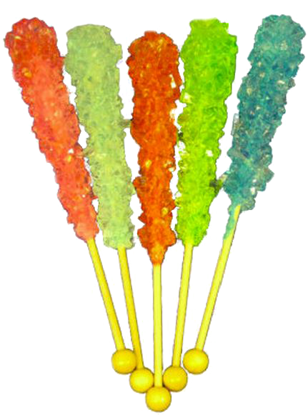
That’s about as easy to do as it sounds, but think of it like one of those rock lollipops that you make by forming sugar crystals on a stick. Same thing really, only with more X-rays and less sugar.


Connect with us on social media